A substantial stride made by AI (Particularly Deep learning) allows the machine to represent complex data. AI is the superset of deep learning, represents the combinational concepts of neurons. Deep learning has achieved strong momentum in the healthcare domain, such as orthopaedic and traumatology. It detects and classifies a different kind of bone fracture using CT scans and X-ray images.
Disability-adjusted life years (DALY) statistics tabulate the number of healthy years lost due to injury, illness, and premature death. By accurate tally and tracking the effect of diseases, DALY’s have opened a new era in health care. Now, vehicular accidents along with heart diseases and depressions are among the top three sources of DALY’s. An increase in the number of vehicles and more travel to fulfil the different life-related issues has increased vehicular accidents. Bone injuries and fractures are the main health-related issues due to vehicular accidents in day-to-day life.
Different types of bone fractures are caused by traumas or specific bone diseases and macro fractures in healthy bone. Microfractures are known as “Stress fractures”. These micro-fractures normally occur after continuous loading, which ultimately may result in a macro fracture. Inducing stresses by the accidental load on a bone causes “Traumatic fracture”. Bone having inferior mechanical properties are more susceptible to traumatic fractures. Some bone diseases (neoplasm, malnutrition, etc.) cause bone destruction or weakening may produce a fracture called “Pathogenic fracture”. Further, bone fractures can also be classified based on the shape and pattern of fractured fragments. They may be transverse, oblique, spiral, and comminuted. Crush fractures, gunshot fractures, greenstick fractures, and avulsion are other types of fractures. Based on aetiology, it can be divided into traumatic, fatigue, and pathological fractures.
Fractures healing is an important biological method necessary for the fitness of injured animals/humans. Regaining the initial bone strength is of great biological importance. Three main steps are involved in the healing/repair process of bone – inflammatory stage, fibroplasia stage, and remodelling stage. Fracture healing can be classified as direct and indirect healing. “Direct healing” is a faster process. It involves intra membranous bone formation and direct cortical remodelling. Osteons can cross the fracture site and bridge the gap, which lays down the cylinders of bone, and the fractures are healed with the help of numerous osteons. It takes a few months to repair. “Indirect fracture healing” is an ordered process of bone repair and reorganization. Steps in indirect healing are impaction, inflammation, soft callus formation, callus mineralization, and callus remodelling.
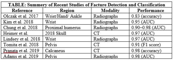
A bone fracture can be diagnosed based on history and physical examination. X-ray, CT scan, or an MRI are used to view the suspected bone fracture. In medical application sensitivity and accuracy of medical problem, detections have two measures. Nowadays, artificial intelligence (AI) has recently made a more sensitive and accurate role in detecting a health problem through machines, as shown in the table.

The use of AI in orthopaedics and traumatology to detect fractures is an emerging tool to save time, accurate, sensitive detection of fracture, and measurement.

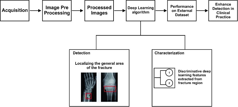
Kalmet et al., (2020) suggested deep learning (DL) methods. According to them, DL is a part of a broad machine learning field and a broader AI field. Algorithms used in this process can be able to solve various problems in different quantitative fields. AI can be used in fracture detection and fracture characterization tasks, which will improve the speed and accuracy of fracture diagnosis, as shown in the figure.
As we know machine/deep learning is a resource-hungry job, which means that the more we train the algorithm, testing results will also be better and accurate. So that, a well-trained algorithm can reduce the workforce of the human radiologist. Deep learning also applied to radionics, which extracts the quantitative features from particular medical images that cannot be accessible by the human eyes.
AI can expand their library for clinical applications. Several promising studies explain the improved performance of AI in clinical tasks like fracture detection on X-ray and CT-Scan. _______________________________________________________
References
Adams M, Chen W, Holcdorf D, McCusker M W, Howe P D, Gaillard F. Computer vs human: deep learning versus perceptual training for the detection of neck of femur fractures. J Med Imaging Radiat Oncol 2019; 63: 27-32.
Chung S W, Han S S, Lee J W, Oh K S, Kim N R, Yoon J P, Kim J Y, Moon S H, Kwon J, Lee H J, Noh Y M, Kim Y. Automated detection and classification of the proximal humerus fracture by using deep learning algorithm. Acta Orthop 2018; 89: 468-73.
Heimer J, Thali M J, Ebert L. Classification based on the presence of skull fractures on curved maximum intensity skull projections by means of deep learning. J Forensic Radiol Imaging 2018; 14: 16-20.
Kalmet, Pishtiwan HS, et al. “Deep learning in fracture detection: a narrative review.” Acta orthopaedica 91.2 (2020): 215-220.
Kim D H, MacKinnon T. Artificial intelligence in fracture detection: transfer learning from deep convolutional neural networks. Clin Radiol 2018; 73: 439-45.
Lindsey R, Daluiski A, Chopra S, Lachapelle A, Mozer M, Sicular S, Hanel D, Gardner M, Gupta A, Hotchkiss R, Potter H. Deep neural network improves fracture detection by clinicians. Proc Natl Acad Sci USA 2018; 115: 11591-6.
Olczak J, Fahlberg N, Maki A, Razavian A S, Jilert A, Stark A, Sköldenberg O, Gordon M. Artificial intelligence for analyzing orthopedic trauma radiographs. Acta Orthop 2017; 88: 581-6.
Pranata Y D, Wang K C, Wang J C, Idram I, Lai J Y, Liu J W, Hsieh I H. Deep learning and SURF for automated classification and detection of calcaneus fractures in CT images. Comput Methods Programs Biomed 2019; 171: 27-37.
Tomita N, Cheung YY, Hassanpour S. Deep neural networks for automatic detection of osteoporotic vertebral fractures on CT scans. Comput Biol Med 2018; 98: 8-15.


